Research on Brain Cell Development
This page is dedicated to the families who made the difficult choice to support brain research by donating their loved ones' brain tissue. Postmortem research is essential in discovering microstructural brain differences in autism and this research is not possible without the strength and foresight of these incredible families.
We hope that, in some small way, our research brings meaning to your loss. Thank you.
Brain tissue research
 To explore the brain-based cause and manifestation of autism, it is necessary to look at postmortem brain tissue. Doing so gives us a first-hand view of the changes in brain cells (neurons):
- if they are misshapen
- if they are located in incorrect regions
- if there are too many/too few of them
Brain growth linked with excess cells counts
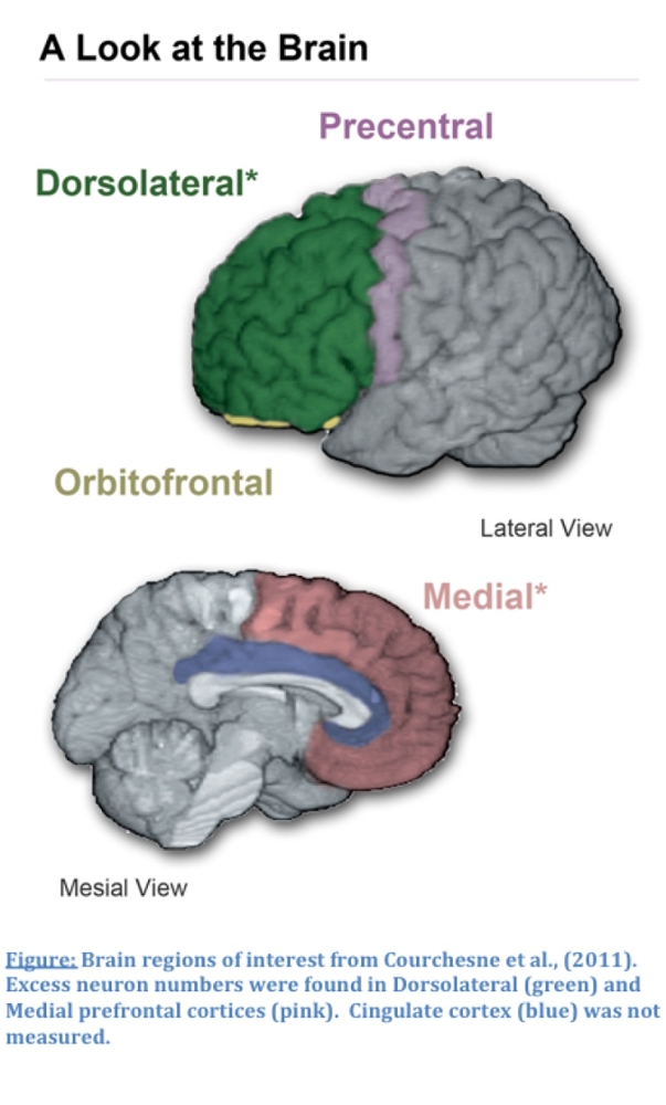
The Autism Center of Excellence has identified a possible driver for brain overgrowth in boys with autism. In a study published in the Journal of the American Medical Association (see the abstract at Courchesne and colleagues, 2011), we discovered a 67% excess in the number of neurons found in brain regions of boys with autism associated with social, communication and cognitive development. Co-investigator Peter Mouton was able to identify this excess cell count using blinded (un-biased) stereological methods. This finding suggests a potential cause for the rapid change in growth trajectories observed in autistic infants toddlers. Understanding the impact of the increased neuron numbers on development in autistic children will provide additional insight into the actual brain-based mechanisms underpinning the disorder.
Next steps: Understanding why and what impact more cells will have
The ACE is actively involved in exploring this finding with the help of our collaborators. Conducting postmortem research is exceptionally challenging and requires a skilled team of researchers, capable institutes, and agencies to provide high quality research materials. Leading the next steps of this research are ACE Director Eric Courchesne, Ed Lein at the Allen Institute for Brain Sciences, Sophia Colamarino, and Tony Wynshaw-Boris. None of this work would be possible without the great resources provided by the NIH Developmental Brain and Tissue Bank and the Autism BrainNet (formerly known as the Autism Tissue Program).
Related Publications
-
Courchesne E, Mouton PR, Calhoun ME, Semendeferi K, Ahrens-Barbeau C, Hallet MJ, Barnes CC, Pierce K. Neuron number and size in prefrontal cortex of children with autism. JAMA. 2011 Nov 9;306(18):2001-10. PubMed PMID: 22068992.
-
Chow ML, Li HR, Winn ME, April C, Barnes CC, Wynshaw-Boris A, Fan JB, Fu XD, Courchesne E, Schork NJ. Genome-wide expression assay comparison across frozen and fixed postmortem brain tissue samples. BMC Genomics. 2011 Sep 10;12:449. PubMed PMID: 21906392; PubMed Central PMCID: PMC3179967.
-
Kennedy DP, Semendeferi K, Courchesne E. No reduction of spindle neuron number in fronto-insular cortex in autism. Brain Cogn. 2007 Jul;64(2):124-9. Epub 2007 Mar 13. PubMed PMID: 17353073.
-
Buxhoeveden DP, Semendeferi K, Buckwalter J, Schenker N, Switzer R, Courchesne E. Reduced minicolumns in the frontal cortex of patients with autism2. Neuropathol Appl Neurobiol. 2006 Oct;32(5):483-91. Erratum in: Neuropathol Appl Neurobiol. 2007 Dec;33(6):720-1. Neuropathol Appl Neurobiol. 2007 Oct;33(5):597. PubMed PMID: 16972882.
- Courchesne E, Redcay E, Morgan JT, Kennedy DP. Autism at the beginning: microstructural and growth abnormalities underlying the cognitive and behavioral phenotype of autism. Dev Psychopathol. 2005 Summer;17(3):577-97. Review. PubMed PMID: 16262983.
