
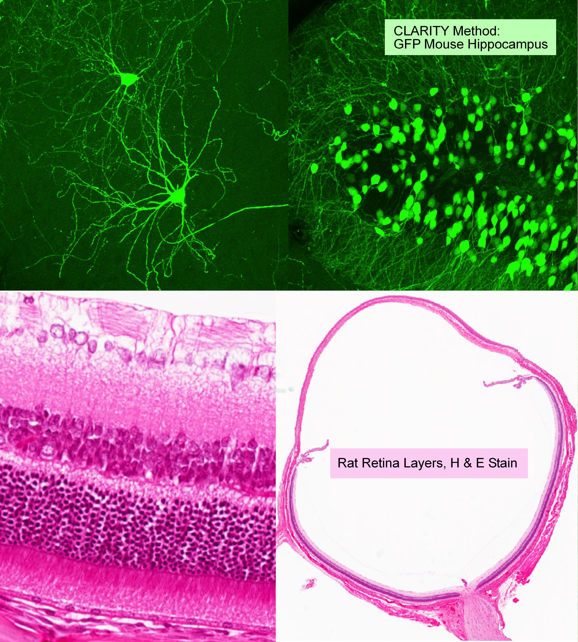
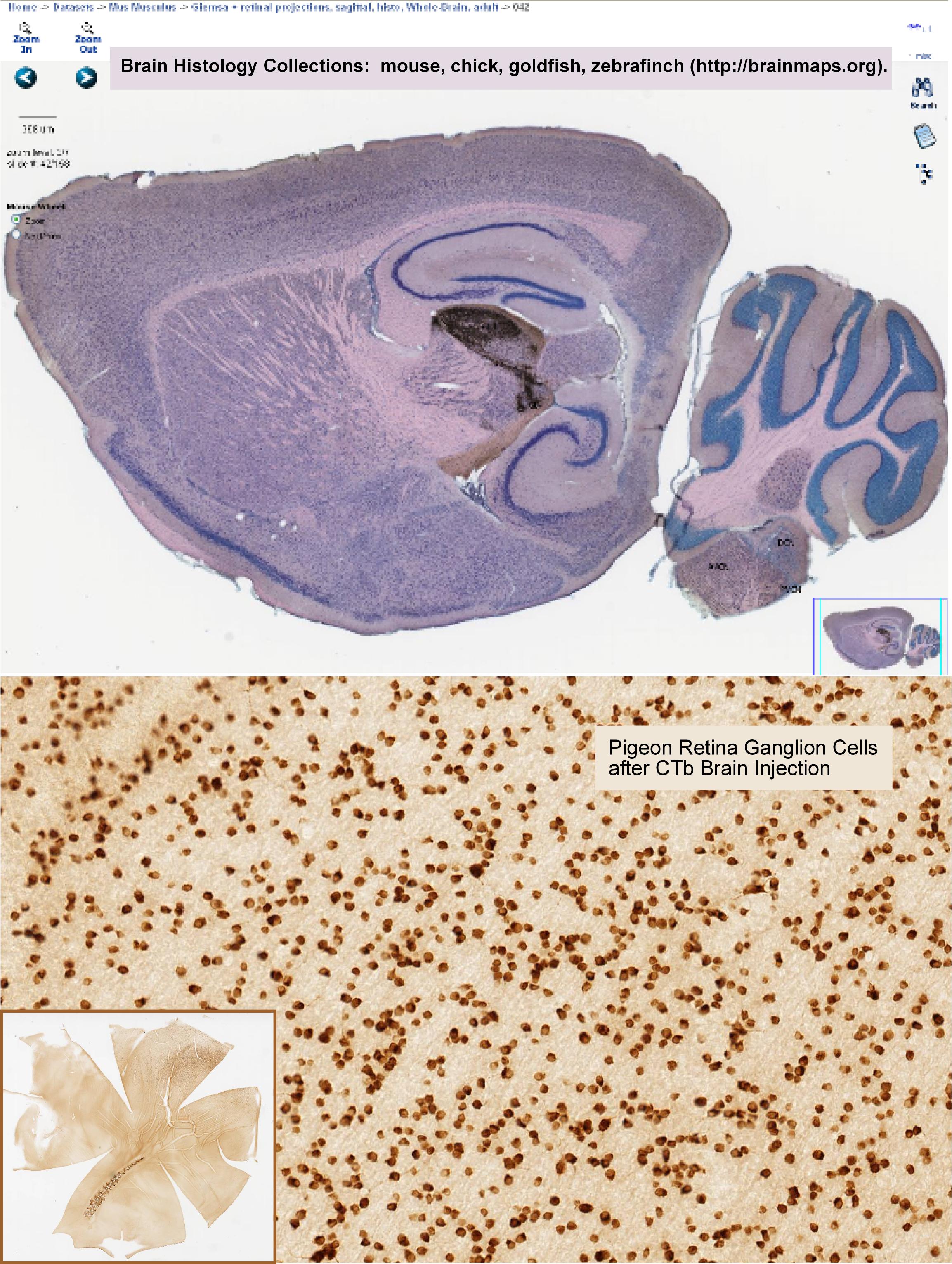
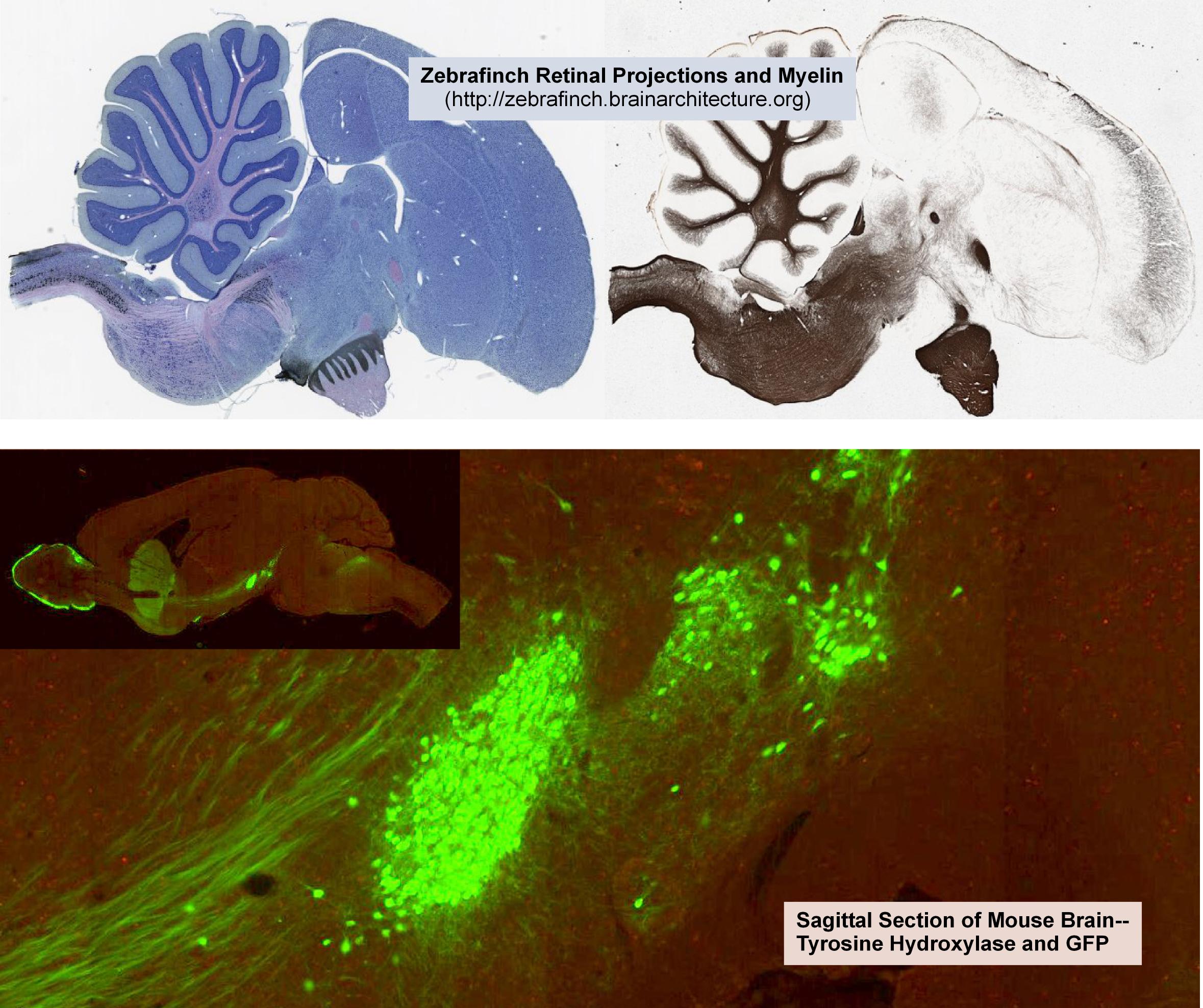
Histology Images
Slide Scanning is an ideal method for imaging whole tissue sections. The Core has a Hamamatsu slide scanner for fluroescence and brightfield.
The Histology services offered by our Core do not include parrafin or OCT embedding, sectioning and staining. We only perform electron microscopy preparations for serial block face sectioning, clearing of tissues, and array tomography.
Samples of slide scanning images:
Citation Guidelines for Using the SOM Microscopy Core
All publications using SOM Microscopy Core resources must cite the following grant numbers:
NS047101, OD030505, OD036455
Example acknowledgment:
Microscopy imaging was performed at the UCSD SOM Microscopy Core
(NS047101, OD030505, OD036455).