
Jacob Koffler, Ph.D., MBA
Assistant Professor

- Neural Engineering Lab
- Publications
Neural Engineering Lab
Our lab is interested in applying bioengineering approaches to promote regeneration in the central and peripheral nerve regeneration. We use 3D printing, stem cells, material sciences, and drug delivery to provide guidance and enhance axonal regeneration.
Our scaffold design is inspired by nature; thus, we fabricate biomimetic structures that can support improved regeneration. Our scaffolds enable topographic patterning of regenerating axons in order to guide them into their respective fascicles, thus re-aligning them to their respective targets. Using linear micro-channels that restrict axonal regeneration to one direction, we prevent axonal probing of the 3D lesion space (as happens in cellular grafts). Thus, axons do not regenerate randomly in all directions, rather they bridge the injury site and reach the same respective tract.
We use these scaffolds to study axonal regeneration in models of traumatic spinal cord injury and peripheral nerve injury.
In the context of spinal cord injury we also load scaffolds with neural stem cells that form a neuronal relay between host regenerating axons through the lesion site and into the caudal intact spinal cord.
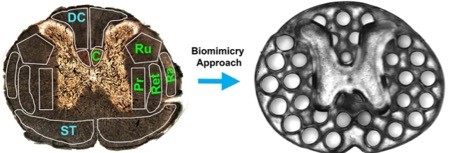
Axonal projections in the spinal cord are linearly organized into regions (fascicles) containing axons of related function. Motor systems are shown in green and sensory systems in blue. C, Corticospinal tract. Ru, Rubrospinal tract; Ra, Raphespinal tract; Ret,Reticulospinal tract; Pr, Propriospinal tract; ST, Spinothalamic tract; DC, dorsal column sensory axons. Channels are precisely printed in 3D space. Collaboration with Dr. Shaochen Chen, Deprtment of Nanoengineering, UCSD.
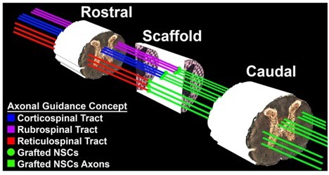
Hypothetical alignment and guidance of regenerating host axons and stem cell axons: Scaffold walls confine both host and stem cell axon growth to linear rostral-caudal planes. Thus, a lesioned host axon (e.g. CST, Rubro or Reticulospinal axons) regenerates into the scaffold and forms a synapse onto a stem cell neuron inside a channel, and the stem cell neuron in turn extends an axon out of the scaffold below the injury site (green lines) into the same white matter fasciculus below the lesion, guided by the microchannel architecture of the scaffold. The scaffold maintains the exact 3D coordinates throughout the lesion site, matching natural host architecture.
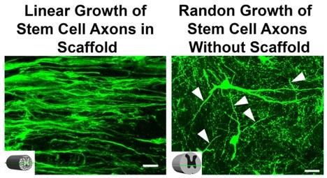
Stem Cell Derived-Axons are Guided by 3D Printed Microchannel Scaffold.
Rat GFP-expressing neural stem cells loaded in 3D printed PEG/GelMa scaffold. Stem cell-derived axons within channels extend in linear configurations along rostral-to-caudal axis of the lesion site after one month in vivo, whereas neural stem cells implanted as a cell suspension into the lesion site, without a scaffold, extend axons in random orientations in the lesion site (arrowheads). The schematic indicates the orientation of the horizontal section. Scale bar: 40µm
In the context of peripheral nerve injury we implant our scaffolds in models sciatic nerve transection.
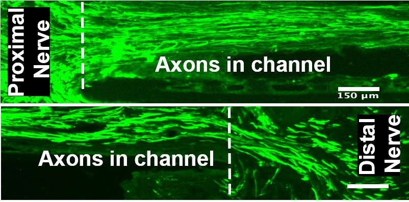
Scaffolds with strictly linear internal architecture successfully guide and enhance axonal regeneration into and beyond sites of peripheral nerve injury.
Publications
1. Koffler J.*, Zhu W, Qu X, Platoshyn O, Dulin J, Brock J, Graham L, Lu P, Sakamoto J, Marsala M, Chen S, Tuszynski MH. Biomimetic 3D Printed Spinal Cord Scaffolds for Spinal Cord Injury. Nature Medicine. 2019Upd Jan 14 . * Corresponding Author.
2. Pawelec KM*, Koffler J.*, Shahriari D, Galvan A, Tuszynski MH, Sakamoto J. Microstructure and in vivo Characterization of Multi-channel Nerve Guidance Scaffolds. Biomed Mater. 2018 Feb 7. * co-first authors.
3. Shahriari D, Shibayama M, Lynam D, Wolf K, Kubota G, Koffler J., Tuszynski M, Campana W, Sakamoto J. Peripheral Nerve Growth within a Hydrogel Microchannel Scaffold Supported by a Kink-Resistant Conduit. J Biomed Mater Res A. 2017 Aug 14.
4. Shahriari D, Koffler J., Tuszynski MH, Campana WM, Sakamoto JS. Hierarchically Ordered Porous and High-Volume Polycaprolactone Microchannel Scaffolds Enhanced Axon Growth in Transected Spinal Cords. Tissue Eng Part A. 2017 May;23(9-10):415-425.
5. Shahriari D, Koffler J., Lynam DA, Tuszynski MH, Sakamoto JS. Characterizing the degradation of alginate hydrogel for use in multilumen scaffolds for spinal cord repair. J Biomed Mater Res A. 2016 Mar;104(3):611-619.
6. Brock JH, Graham L., Staufenberg E., Collyer E., Koffler J., MH Tuszynski. Bone Marrow Stromal Cell Transplants Fail to Improve Motor Outcomes in a Severe Model of Spinal Cord Injury. J Neurotrauma. 2016 Jun 15;33(12):1103-14.
7. Lynam DA, Shahriari D., Felger KJ, Angart PA, Koffler J., Tuszynski MH, Chan C., Walton P., Sakamoto J. Brain Derived Neurotrophic Factor Release from Layer-by-Layer Coated Agarose Nerve Guidance Scaffolds. 2015 Acta Biomaterialia, Accepted.
8. Shandalov Y.*, Egozi D.*, Koffler J., Dado-Rosenfeld D., Ben-Shimol D., Shor E., Kabala A., Levenberg S., An engineered muscle flap for reconstruction of large soft tissue defects, Proc Natl Acad Sci U S A., 2014 April 7.
9. Koffler J., Samara R. Rosenzweig E. Invited chapter: Using Templated Agarose Scaffolds to Promote Axon Regeneration Through Sites of Spinal Cord Injury. Methods Mol Biol. 2014;1162:157-65.
10. Kaufman-Francis K, Koffler J., Weinberg N, Dor Y, Levenberg S, Engineered Vascular Beds Provide Key Signals to Pancreatic Hormone-Producing Cells. (2012) PLoS ONE 7(7): e40741
11. Koffler J., Kaufman-Francis K., Shandalov Y., Egozi D., Amiad-Pavlov D., Landesberg A., Levenberg S. Improved vascular organization enhances functional integration of engineered skeletal muscle grafts. Proc Natl Acad Sci U S A. 2011 Sep 6;108(36):14789-94
13. Lesman A., Koffler J., Atlas A., Blinder Y., Kam Z., Levenberg S. Engineering Vessel-Like Networks Within Multicellular Fibrin-Based Constructs. Biomaterials. 2011 Nov;32(31):7856-69.
13. Rivkin I., Choen K., Koffler J., Melikhov D., Peer D., Margalit R. Paclitaxel-clusters coated with hyaluronan as selective tumor-targeted nanovectors. Biomaterials 31 (2010) 7106e-7114
