
Research Agenda
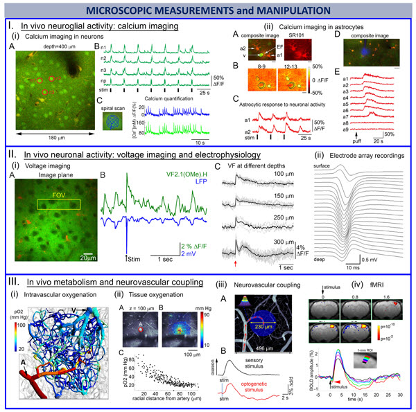
We apply a suite of 2-photon imaging methods to quantify differential vasoactive role, energetic costs, and electrical currents/potentials associated with activity of different neuronal cell types. The goal of these experiments is to determine how activity of a particular type of neurons affects CBF, CMRO2, and current dipole. Our 2-photon methods include high-resolution longitudinal measurements on neuronal activity (Ii, IIi), astrocytic reactivity/calcium surges (Iii), intravascular/tissue oxygen availability and consumption (IIIi-ii), and others. These measurements are combined with laminar and surface electrophysiological recordings (IIii). Selective stimulation of specific neuronal cell types is achieved using optogenetics (IIIiii), both in optical and fMRI experiments (IIIiv). Mouse fMRI provides a translational bridge and is used for validation of the bottom-up modeling effort.
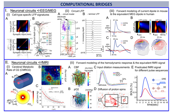
Experimentally obtained microscopic parameters is used for “bottom-up” simulation of macroscopic CBF and CMRO2, that can be obtained noninvasively using calibrated BOLD fMRI, and current dipole moment, that can be obtained noninvasively using MEG. Top row: Existing knowledge about the projection domains of different types of inhibitory neurons segregating along Pyramidal cells projection domains of different types of inhibitory neurons producing dipoles (Ii). Their summed contributions can be detected by laminar recordings of extracellular potentials (Iii) that can be used to forward calculate MEG signals (Iiii). Bottom row: Knowing microscopic distributions of CBF and CMRO2, we can calculate the corresponding BOLD fMRI signal.
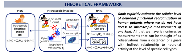
The overarching idea is that the activation of different neuronal cell types has different signatures in the evoked CBF (ΔF), CMRO2 (ΔC), and the macroscopic current dipole responses (ΔD), and by measuring these responses noninvasively in the human brain we will be able to probe more deeply the underlying cellular and circuit activity. For each neuronal population (e.g., excitatory and inhibitory), the current dipole moment (measured by MEG) and CBF/CMRO2 (measured by calibrated BOLD) can be expressed as convolution of the neuronal activity for neuronal population of cell type j with time-resolved weighting factor W(t) for that cell type (that can be thought of as cell-type-specific impulse response function, IRF). The weighting factors W(t) are determined experimentally using selective optogenetic activation of specific cell types in mice.