Leica Center of Excellence at the University of California San Diego
Leica Microsystems and the UCSD School of Medicine Microscopy Core have partnered to offer researchers in the San Diego area access to cutting-edge instrumentation and expertise to further research initiatives. Established in 2019, this non-exclusive collaboration was founded on a desire to directly connect current research needs with technology development in the field of microscopy. Leica Microsystems has been pivotal at providing support for local research through informational seminars, advanced imaging workshops, and direct access to application specialists and engineers. This collaboration allows the UCSD School of Medicine Microscopy Core to further microscopy research on campus and locally.
The Science Lab is a knowledge portal of Leica Microsystems offers scientific research and teaching material on the subjects of microscopy. The content is designed to support beginners, experienced practitioners and scientists alike in their everyday work and experiments. Explore interactive tutorials and application notes, discover the basics of microscopy as well as high-end technologies – become part of the Science Lab community and share your expertise!
About the UCSD School of Medicine Microscopy Core
The UCSD School of Medicine Microscopy core was established in 2004 to provide researchers with easy and reliable access to instrumentation that individual labs could not afford to purchase and manage. The core has since amassed 11 high end optical instruments and one electron microscope along with software for 3D and 4D analysis. All resources are available to researchers and students along with training and guidance. The core is supervised by two expertly trained and experienced microscopists who provide on demand technical support and research advice. With instrumentation and support staff all located in one continuous space in the heart of the School of Medicine, the Microscopy Core has become a staple for imaging research at UCSD with over 700 publications and over 200 laboratories actively utilizing core resources.
Available Resources at the School of Medicine Microscopy Core
Instrumentation representing the Leica Center of Excellence
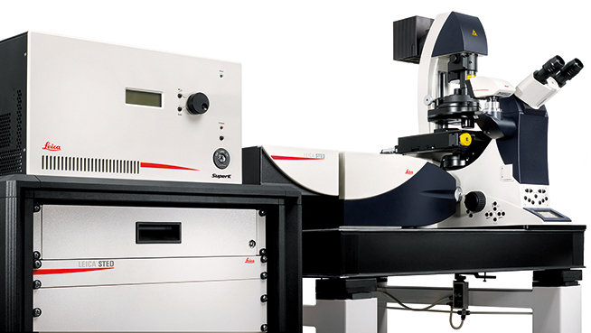
Leica SP8 Confocal with White Light Laser, STED and FALCON
Leica SP8 Confocal with White Light Laser, STED and FALCON

What it’s good for
- STED Super Resolution <50nm in XY
- Tunable excitation source for hand picking excitation wavelengths of up to 8 simultaneously (470nm-670nm)
- FLIM acquisition with Falcon module and Phasor Plot
- Incubation for live cell imaging (BSL1 and BSL2 cell lines)
- Determining colocalization for multiple signals
- Acquisition of up to 5 color channels simultaneously
- Acquisition of multiple color channels sequentially
- Acquisition of a transmitted light image with DIC
- Fixed cells or tissues on a slide with a coverslip (#1.5) or in a dish
- Spectral Deconvolution to correct for overlapping signals
- On the fly Lightning image deconvolution for super resolution of 120nm in X and Y and 200nm in Z
Principles of operation
Laser light of specific wavelengths is scanned across the sample and filtered before detection to produce a high resolution image composed of a small optical slice of the sample. STED enahances resolution by shrinking fluorescence to a small spot with a depletion laser. Deconvolution further enhances resolution, including STED images. FALCON measures time domain fluorescence lifetime values. Phasor plots time domain fluorescence lifetime values for easy visualization.
Technical information
- Microscope: Leica DMi8 Inverted
- Spectral emission collection up to 800nm
- 2 PMT and 3 HyD detectors
- STED - 3 Super Resolution depletion lines
- Pulsed White Light Laser tunable to any desired wavelength between 470 and 670nm
- 440nm pulsed laser for FLIM
- 405nm laser
- 10x (.40 NA)
- 20x (.75 NA)
- 40x Oil (1.30 NA)
- 40x Water (1.10 NA)
- 63x Oil (1.40 NA)
- 100x Oil (STED)(1.40 NA)
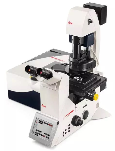
Leica SP8 Confocal
Leica SP8 Confocal

What it’s good for
- Super Resolution Confocality
- Determining colocalization for multiple signals
- Acquisition of up to 4 color channels simultaneously, 2PMTs and 2 HyDs
- Acquisition of multiple color channels sequentially
- Acquisition of a transmitted light image with DIC
- Fixed cells or tissues on a slide with a coverslip (#1.5) or in a dish
- Spectral Deconvolution to correct for overlapping signals
- On the fly Lightning image deconvolution for super resolution of 120nm in X and Y and 200nm in Z
What it’s not good for
- Live Cells: This system does not have incubation for live cell applications
Principles of operation
Laser light of specific wavelengths is scanned across the sample and filtered before detection to produce a high resolution image composed of a small optical slice of the sample. Deconvolution further enhances resolution.
Technical information
- Microscope: Leica DMi8 Inverted
- Spectral emission for all channels
- 405nm
- 488nm
- 552nm
- 638nm
- 10x (.40 NA)
- 20x (.75 NA)
- 40x Oil (1.30 NA)
- 63x Oil (1.40 NA)
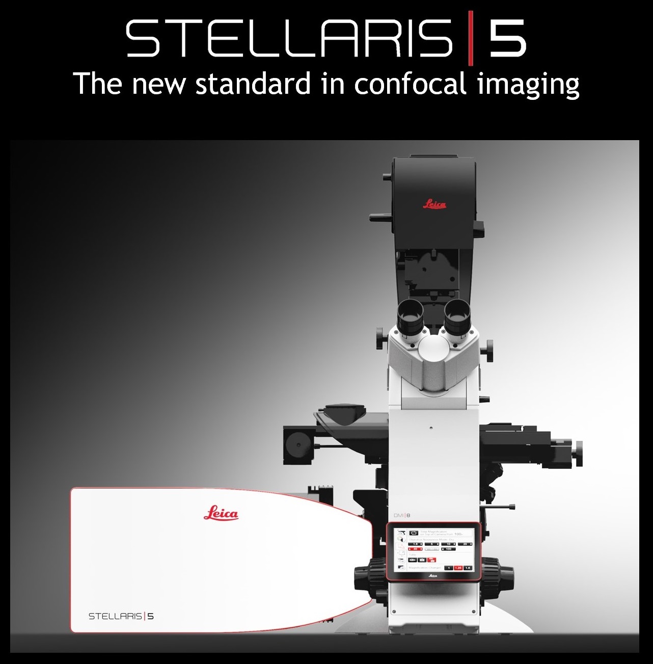
Leica Stellaris 5 Confocal with White Light Laser
Leica Stellaris 5 Confocal with White Light Laser

What it’s good for
- Super Resolution Confocality
- Tunable excitation source for hand picking excitation wavelengths of up to 8 simultaneously
- Determining colocalization for multiple signals
- Acquisition of up to 5 color channels simultaneously, all HyDs
- Acquisition of multiple color channels sequentially
- Acquisition of a transmitted light image with DIC
- Fixed cells or tissues on a slide with a coverslip (#1.5)
- Spectral Deconvolution to correct for overlapping signals
- On the fly Lightning image deconvolution for super resolution of 120nm in X and Y and 200nm in Z
- TauSense tools for fast FLIM and Gating
What it’s not good for
- Live Cells: This system does not have incubation for live cell applications
- Samples in a dish or plate
Principles of operation
Laser light of specific wavelengths is scanned across the sample and filtered before detection to produce a high resolution image composed of a small optical slice of the sample. Deconvolution further enhances resolution.
Technical Information
- Microscope: Leica DMi8 Inverted
- Spectral emission for all channels
- White Light Laser (470nm - 790nm)
- 5x (.15 NA)
- 10x (.40 NA)
- 20x Oil/Water/Glycerol (.75 NA)
- 20x (.40 NA)
- 40x Oil (1.30 NA)
- 63x Oil (1.40 NA)
