Events & Updates
Coming Soon
New Microscopy Core Resources
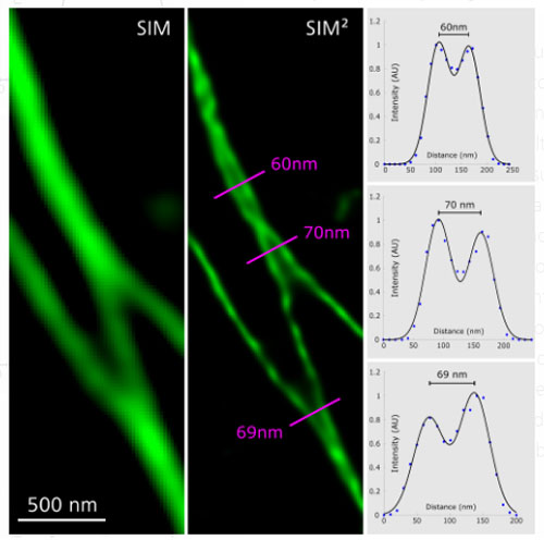
Zeiss Elyra 7 with Lattice SIM
- Resolve structures down to 60 nm.
- Observe live cell dynamics at up to 255 fps.
- Accelerate image acquisition in all three dimensions.
- Get the sharpest sectioning in wide-field microscopy.
- Utilize a wealth of imaging techniques on one platform.
Contact us today for a free demonstration: jsantini@health.ucsd.edu

Leica Center of Excellence
The UC San Diego School of Medicine Microscopy Core is now a Leica Center of Excellence! We have three Leica systems that are supported by Leica:
- Leica SP8 Confocal with Lightning Deconvolution
- Leica SP8 Confocal with White Light Laser, Falcon (FLIM), STED and Lightning Deconvolution
- Leica Stellaris 5 Confocal with Lightning Deconvolution and TauSense Tools
If you would like to benefit from our center's collaboration with Leica, you can do so in the following ways:
Write a White Paper!
White papers are short descriptions of your research application. Descriptions include use of our Leica systems along with a beautiful image.
Write a Techical Note!
Technical notes consist of more indepth description of methods used or developed on the Leica platform.
Both white papers and technical notes are a great way for you to increase the impact of your publication! Readers will naturally visit your publication after reading your Leica collaboration and potentially cite your paper.
Give a Webinar!
Leica can promote your lab and your research through hosting your talk to the world!
Be a guest speaker at a local conference!
Leica can host you to be a guest speaker at one of the many local conferences held in San Diego.
If you are interested in any of these options or would like more information, please email us for details.
Webinars, Seminars and Demonstrations
Citation Guidelines for Using the SOM Microscopy Core
All publications using SOM Microscopy Core resources must cite the following grant numbers:
NS047101, OD030505, OD036455
Example acknowledgment:
Microscopy imaging was performed at the UCSD SOM Microscopy Core
(NS047101, OD030505, OD036455).
