
Resources & Services
Training: To arrange training on any resources, contact Jennifer Santini, Managing Director.
Microscopes
NEW! - Evident FV5000 Multiphoton Microscope
What it’s good for
- Multiphoton deep tissue imaging and live animal imaging
- Confocal Microscopy
- Tunable Multiphoton excitation source from 700-1300nm and fixed excitation at 1040nm
- Fixed single photon excitation lines (405, 488, 561, 635)
- Determining colocalization for multiple signals
- Second and Third Harmonic Generation for label free imaging
- Acquisition of up to 4 color channels simultaneously with Multiphoton
- Acquisition of up to 2 color channels simultaneously with Confocal
- Acquisition of multiple color channels sequentially with Multiphoton and Confocal
- Acquisition of a transmitted light image
What it’s not good for
- Live Cells: This is an upright system with no incubation
- Samples in a plate
Principles of operation
Laser light of specific wavelengths is scanned across the sample and filtered before detection to produce a high resolution image composed of a small optical slice of the sample.
Technical Information
Multiple confocal objectives available for magnification and resolution
Multiphoton Objective:
- 25x 1.00NA
- 2mm working distance
- adjustable correction collar
Olympus VS200 Slide Scanner - Fluorescence and Brightfield
What it's good for
- Generating brightfield and multi-color fluorescent images of entire slides or tissue sections automatically
- 7 fluorescent colors
- Fixed cells or sections on a standard 1 x 3 slide with a coverslip (#1.5) or 2 x 3 slides
- Can scan up to 210 slides automatically
What it's NOT good for
- Thick tissue slices (greater than 50um)
- Timelapse or live cell applications
Principles of operation
Fluorescent light from an LED lamp filtered for emission or brighfield light produce an image by tiling specified areas of a slide to generate montage reconstructions.
Technical information
- 7 fluorescent color channels and brightfield:
- DAPI - Ex 345 Em 432/36
- FITC - Ex 494 Em 515/30
- TRITC - Ex 555 Em 600/31
- CY5 - Ex 625 Em 685/42
- Cy7 - Ex 743 Em 810/81
- CFP - Ex 458 Em 482/25
- YFP - Ex 513 Em 544/25
- Motorized X-Y capability for slide scanning
- Microscope objective:
- 2x, 4x, 20x (.80 NA), 40x Air
Files must be named in the proper format to open. There is a folder and thumbnail for each image.
Example
For slide preparations
- No wet slides - must be completely dry
- Use #1 or #1.5 coverslips
Software for Processing
NEW! - Olympus VS200 Slide Scanner - Brightfield
What it's good for
- Generating brightfield images of entire slides or tissue sections automatically
- 7 fluorescent colors
- Fixed cells or sections on a standard 1 x 3 slide with a coverslip (#1.5) or 2 x 3 slides
- Can scan up to 210 (1 x 3) or 15 (2 x 3) slides automatically
What it's NOT good for
- Thick tissue slices (greater than 50um)
- Timelapse or live cell applications
Principles of operation
Fluorescent light from an LED lamp filtered for emission or brighfield light produce an image by tiling specified areas of a slide to generate montage reconstructions.
Technical information
- Motorized X-Y capability for slide scanning
- Microscope objective:
- 2x, 4x, 20x (.80 NA), 40x air
Files must be named in the proper format to open. There is a folder and thumbnail for each image.
Example
For slide preparations
- No wet slides - must be completely dry
- Use #1 or #1.5 coverslips
Software for Processing
Leica Stellaris 5 Confocal with White Light Laser
What it’s good for
- Super Resolution Confocality
- Tunable excitation source for hand picking excitation wavelengths of up to 8 simultaneously
- Determining colocalization for multiple signals
- Acquisition of up to 5 color channels simultaneously, all HyDs
- Acquisition of multiple color channels sequentially
- Acquisition of a transmitted light image with DIC
- Fixed cells or tissues on a slide with a coverslip (#1.5)
- Spectral Deconvolution to correct for overlapping signals
- On the fly Lightning image deconvolution for super resolution of 120nm in X and Y and 200nm in Z
- TauSense tools for fast FLIM and Gating
What it’s not good for
- Live Cells: This system does not have incubation for live cell applications
- Samples in a dish or plate
Principles of operation
Laser light of specific wavelengths is scanned across the sample and filtered before detection to produce a high resolution image composed of a small optical slice of the sample. Deconvolution further enhances resolution.
Technical Information
White Light Laser (470nm - 790nm)
- 5x (.15 NA)
- 10x (.40 NA)
- 20x Oil/Water/Glycerol (.75 NA)
- 20x (.40 NA)
- 40x Oil (1.30 NA)
- 63x Oil (1.40 NA)
Leica SP8 Confocal with Lightning Deconvolution
What it’s good for
- Super Resolution Confocality
- Determining colocalization for multiple signals
- Acquisition of up to 4 color channels simultaneously
- Acquisition of multiple color channels sequentially
- Acquisition of a transmitted light image with DIC
- Fixed cells or tissues on a slide with a coverslip (#1.5) or in a dish
- Spectral Deconvolution to correct for overlapping signals
- On the fly image deconvolution for super resolution of 120nm in X and Y and 200nm in Z
What it’s not good for
- Live Cells: This system does not have incubation for live cell applications
Principles of operation
Laser light of specific wavelengths is scanned across the sample and filtered before detection to produce a high resolution image composed of a small optical slice of the sample. Deconvolution further enhances resolution.
Technical information
- Microscope: Leica DMi8 Inverted
- Spectral for all channels laser lines
- 405nm
- 488nm
- 552nm
- 638nm
Microscope objectives
- 10x (.40 NA)
- 20x (.75 NA)
- 40x Oil (1.30 NA)
- 63x Oil (1.40 NA)
Software & supplemental information
Leica SP8 Confocal with White Light Laser, Falcon (FLIM), STED, and Lightning Deconvolution
What it’s good for
- STED Super Resolution <50nm in XY
- Tunable excitation source for hand picking excitation wavelengths of up to 8 simultaneously (470nm-670nm)
- FLIM acquisition with Falcon module
- Incubation for live cell imaging (only BSL1 cell lines)
- Determining colocalization for multiple signals
- Acquisition of up to 5 color channels simultaneously
- Acquisition of multiple color channels sequentially
- Acquisition of a transmitted light image with DIC
- Fixed cells or tissues on a slide with a coverslip (#1.5) or in a dish
- Spectral Deconvolution to correct for overlapping signals
- On the fly image deconvolution for super resolution of 120nm in X and Y and 200nm in Z
Principles of operation
Laser light of specific wavelengths is scanned across the sample and filtered before detection to produce a high resolution image composed of a small optical slice of the sample. STED enahances resolution by shrinking fluorescence to a small spot with a depletion laser. Deconvolution further enhances resolution, including STED images.
Technical information
- Microscope: Leica DMi8 Inverted
- Spectral for all channels
- PMT and HyD detectors
- STED - Super Resolution depletion lines
- White Light Laser tunable to any desired excitation between 470 and 670nm
Microscope objectives
- 10x (.40 NA)
- 20x (.75 NA)
- 40x Water (1.10 NA)
- 40x Oil (1.30 NA)
- 63x Oil (1.40 NA)
- 100x Oil (STED)(1.40 NA)
Sample preparation guidelines For STED imaging only:
Choice of samples:
- For Live samples: Media should be clear and contain no phenol red or other color additives
- For Fixed Samples: RI of mounting medium should match RI of immersion used (prolong glass or diamond)
- DAPI should be AVOIDED, especially when using green fluorophores
- Autofluorescence should be extremely low
- It's important that the sample doesn't absorb 592nm, 660nm or 775nm
- Use only #1.5 coverglass, including for glass bottom dishes
Recommended Dyes:
- Single color for 592nm depletion line
- DyLight 488 or 514
- Oregon Green 488 or 514
- AlexaFluor 488 or 514
- ATTO 488 or 514
- Single color for 660nm depletion line
- Alexa 532
- ATTO 532 or 550
- TMR/TRITC
- Cy3
- Alexa 555
- Single color for 775nm depletion line
- ATTO 647N
- Alexa 633
- Alexa 594
- ATTO 590
Recommended Fluorescent Proteins:
- eGFP (484nm ex / 592nm depletion)
- EmGFP (487nm ex / 592nm depletion)
- eYFP (514nm ex / 592/660nm depletion)
- Venus (515nm ex / 592/660nm depletion)
- mCitrine (516nm ex / 592/660nm depletion)
- dsRed (558nm ex / 660nm depletion)
- mStrawberry (574nm ex / 660nm depletion)
- mKate2 (588nm ex / 775nm depletion)
- Fluorescent Proteins to AVOID: mCherry, CFP, tagRFP
Software & supplemental information
Leica LAS Lite Software Download
STED Sample preparation guide - Confocal Application Letter (PDF)
STED Sample Prep Oct 18 (PPTX)
LIGHTNING WhitePaper (PDF)
STED 3X Sample Prep 2018.pdf - Powered by Box.html
Zeiss Elyra 7 Lattice SIM
What it's good for
- Generating multi-color fluorescent images
- Live cell imaging - incubation provided
- Super resolution down to 60nm XY
- 4 fluorescent colors - 2 channel simultaneous acquisition (see table)
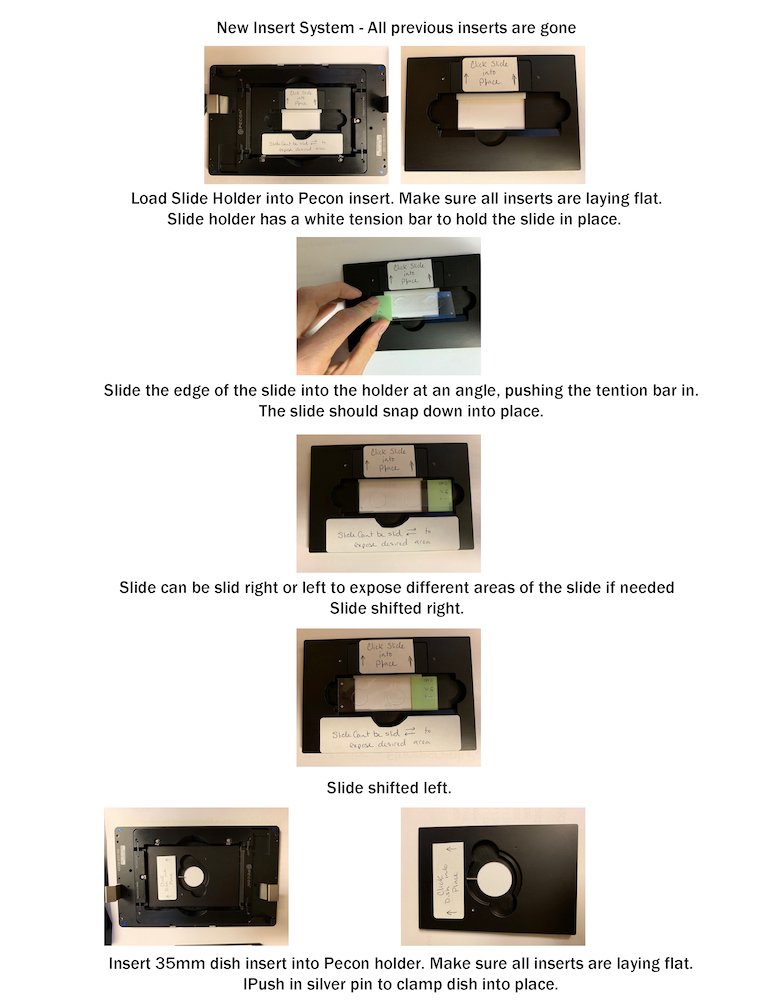
- Fixed cells or sections on a standard 1 x 3 slide with a coverslip (#1.5)
- Live cells in a 35mm glass bottom dish, glass bottom chamber slide, or glass bottom plate
- Apotome imaging
- Lattice structured illumination imaging
- Laser wide-field imaging
- TIRF Imaging
What it's NOT good for
- Thick tissue slices (greater than 100um)
- Lattice SIM is not good for samples with diffuse structureless fluorescence or low contrast
Principles of operation
Fluorescent light from lasers filtered for emission produce an image from wide-field resolutions to super resolutions down to 60nm XY. A lattice pattern is placed in the light path to surpass the diffraction limit and produce super resolutions in lattice SIM. A lattice pattern is placed in the light path to improve resolutions over wide-field.
Technical information
- 4 fluorescent color channels:
- DAPI (405nm)
- FITC (488nm)
- TRITC (561nm)
- CY5 (642nm)
- Motorized X-Y capability for multiposition imaging
- Microscope objectives:
- 10x (.75NA), 20x (.80 NA), 40x (1.40NA water), 63x (1.40NA oil), 100x (1.45NA oil)
Dual Camera Acquisition Parameters:
| Color Combination | Camera 2 | Camera 1 |
| Dapi / TRITC | 420-480nm | 560nm LP |
| Dapi / CY5 | 420-480nm | 655nm LP |
| FITC / TRITC | 495-550nm | 570-620nm |
| FITC / TRITC | 495-525nm | 655nm LP |
For sample preparations
- No wet slides - must be completely dry
- Use #1.5 coverslips or #1.5 coverslip bottom formats
All live cell imaging must be approved by core. Please submit a BUA authorization form along with your lab's BUA report for authorization.
Important Considerations:
- Processing is required when performing Lattice SIM
- A second computer is available for all necessary processing such as SIM2 and Channel Alignment
- Daily alignment of the cameras is required for dual camera acquisition (acquiring two channels simultaneously)
- Alignment of the cameras is not required for single camera use or single fluorophore
- Daily Channel Alignment is required to correct for chromatic shift in XYZ and Rotation when examining more than one fluorophore. To learn about chromatic shift and why it needs to be calibratated the system, please visit:
Gatan 3View SBFS on Zeiss Sigma SEM
System Currently Out of Order
This system is capable of performing the following techniques:
- SEM with high pressure and variable pressure modes
- TEM on grids with STEM detector and 12 grid specimen holder
- Gatan 3View Serial Block Face Scanning Electron Microscopy Demonstration (youtube.com)
- ATLAS software large scale acquisitions of TEM grids and Array Tomography sections
- Correlative Microscopy of Light and Electron microscopy techniques
- Array Tomography with SEM
Principles of operation
Serial block face scanning electron microscopy generates EM resolution 3D images. The system has an ultramicrotome inside the chamber of the SEM, which allows for automatic cutting of a tissue block. The surface of the block is imaged by detection of back-scattered electrons and then a thin section (30-50nm) is cut from the block face. The sample block is then imaged again and a sequence of images can be compiled automatically.
For more information, or access to this technology, contact Jennifer Santini.
Gatan 3View System Publications
Probes for Correlative EM
Colloidal Gold w/wo silver enhancementDAB Photo-oxidation for EM (i.e., Fluorescent Protein, FlAsH and ReAsH, Eosin, miniSOG, APEX, mEos4)
Quantum Dots (size and shape)
En bloc autofluorescence/stains (acridine orange)
Fluorescent Proteins (LR White and LR Gold)
LR White section surface immunofluorescence - Array Tomography (PDF)
Zeiss Z.1 Light Sheet
What it’s good for
- Thick specimens and whole organisms: Cleared tissue, zebrafish etc.
- Fast acquisition speeds and low phototoxicity
- Incubation for live imaging
- Acquisition of up to 2 color channels simultaneously
- Acquisition of multiple color channels sequentially
What it's not good for
- Fixed cells or tissues on a slide with a coverslip (#1.5) or in a dish
Principles of operation
Laser light of specific wavelengths is passed through the sample as a sheet from 1 or 2 sides, perpendicular to the detection optics. Laser excitation, Emission filters and 2 CMOS cameras generate an image at a specific wavelength. Sample is moved through the sheet of light for Z stack acquisition.
Technical information
- Filter based emission for all channels
- 2 pico.edge CMOS cameras
- 3 specimen chambers for different objectives and refractive index
- Incubation for chamber
- Laser Excitations:
- 405nm
- 445nm
- 488nm
- 514nm
- 560nm
- 638nm
Microscope objectives
- EC Plan-Neofluar 5x (.16NA) Air
- CLR Plan-Neofluar 20x Corr (1.0NA) dipping
- Plan-Apochromatic 10x (.5NA) dipping
- W Plan-Apochromatic 20x (1.0NA) dipping
Zeiss LSM 880 Confocal with FAST Airyscan
What it’s good for
- High resolution imaging at 1.7x resolution increase over standard confocal imaging
- High acquisition speed, at 4x faster acquisition than confocal mode
- High sensitivity with use of special detectors
- Shuttle and Find capability to merge light and electron microscopy techniques for Correlative Microscopy
- Acquisition of up to 3 color channels simultaneously
- Acquisition of multiple channels sequentially
- Acquisition of a high contrast DIC image
- Fixed cells or tissues on a slide with a coverslip (#1.5)
- Live cells, small organisms or tissues in a chamber or dish with a coverslip bottom (#1.5) (only BSL1 cell lines)
- Spectral Deconvolution to correct for overlapping signals
- Calcium Dynamics
- FRET
- Stimulus and bleaching applications
What it’s not good for
- Live animal imaging (large animals such as mouse)
Principles of operation
Laser light of specific wavelengths is scanned across the sample and filtered before detection to produce a high resolution image composed of a small optical slice of the sample. Alternative detection at high resolution and sensitivity avoids discrimination of light with a pinhole, and utliizes a 32 detector array that can generate higher resolution images by detection of specific airy units.
Technical information
- Microscope: Ziess Observer inverted stand
- Incubated with CO2
- DIC channel
- laser lines
- 405
- 458
- 488
- 515
- 543
- 594
- 633
MDPI photonics: Exploring the Potential of Airyscan Microscopy for Live Cell Imaging
Zeiss stage inserts
Keyence BZX-700 Fluorescent Microscope
Olympus MVX10 Macroview
What it's good for
- Low magnification observation
- Brightfield (color)
- Darkfield
- Fluorescence
- Obtaining images of:
- intact organs
- whole sections
- Field view from 55mm to 1.74mm
What it's NOT good for
- High magnification
- Multipoint timelapse
- Low contrast brightfield samples
- Low fluorescence
Principles of operation
Fluorescent light from a mercury lamp is filtered for excitation and emission.
Technical information
- Microscope Base: MVX10
- Color CCD camera
- 4 fluorescent color channels:
- DAPI
- FITC
- Texas Red
- Cy5.5
- Color brightfield
- Microscope Objectives:
- .63x
- 2x
Zeiss Upright Widefield Microscope with Apotome for Correlative Array Tomography
Software & Other Resources
Software
Image Pro Plus with 3D Constructor
Powerful 2D and 3D image processing, enhancement, and analysis software with extensive measurement and customization features.
Softworx Suite
Deconvolution with image correction featuring constrained iterative 3D image restoration and image correction, a quantitatively validated deconvolution solution generating the most accurate measure of sample fluorescence available.
Imaris
3D and 4D Real-Time Interactive Image Visualization and Measurements of Large Data Sets, including filament tracing.
Volocity High Performance 3D Imaging Software
High quality and easy to use image processing software with visualization, quantification and restoration Modules for 3D and 4D rendering, measurements and deconvolution.
Free software downloads
- NDP View 2 - Nanozoomer
- Image J LOCI Bioformat Plugin (opening all confocal formats)
- Image J NDPI Tools (for Nanozoomer files)
- Leica LAS Lite Software
- Zen Lite Software from Zeiss
- OlyVia Olympus VS200 Software
Histology Services - Tissue Clearing, EM Sample Preparation and Array Tomography
Services Offered
- CLARITY: Chung, K., Deisseroth, K. CLARITY for mapping the nervous system. Nat Methods 10, 508–513 (2013).
- Tissues successfully cleared so far:
- Brain
- Heart
- Embryos
- Lymph Nodes
- Stomach
- Intestines
- Esophagus
- Spinal Cord
- Optic Nerve
- EM preparations for serial block face sectioning
Services not offered:
- We do not provide standard histology services such as parrafin and OCT embedding and cutting.
- We do not provide staining of cells and tissues.
Array Tomography Information
- The Cellular Scale: Seeing Inside Cells: Array Tomography
- iBiology Techniques YouTube: Microscopy: Array Tomography by Stephan Smith
- Array Tomography with SEM: Serial Section Scanning Electron Microscopy (S3EM) on Silicon Wafers for Ultra-Structural Volume Imaging of Cells and Tissues
Videos for Histological Preparations
Slide Scanning Services
We will scan your slides for you!
Cost
The cost of services includes:
- Prep, load and set up your slide scanning run.
- Hourly microscope charges, plus 1 or more hours of technical assistance fees per session. Time required to perform slide scanning varies based on session type.
Please email us with the following information if you would like to solicit slide scanning services:
Information required for slide scanning services:
- Olympus VS200 (bright-field and fluorescence) or Hamamatsu Nanozoomer (bright-field)
- # of slides to be scanned
- Bright-field or fluorescence
- Sample type – Tissue type / section thickness or monolayer cells mounted on slide
- 1x3 or 2x3 slides
- All fluorescent dyes used in combination
- Full oracle chart string number: Project number - task number - Funding source number
- Bring slides in a well labeled plastic slide box, not a cardboard slide holder
If you want us to do fluorescence scans, you MUST circle the tissue section that you exposure times set from. It should be the first slide in the set. Circle the section in sharpie on the back of the slide, not the coverslip.
Slide scanning software is required to view all slide scanner files:
Olympus VS200 slide scanner (PC only) - OlyVIA V3.3
Hamamatsu Nanozoomer slide scanner (PC and Mac) - NDP.view2
Qpath – Opens all slide scanning files
Safety Requirements
Only BSL1 material is allowed in a majority of the Microscopy Core.
If you wish to do any live cell imaging in the core, you must have approval by submitting a BUA authorization form to core staff along with a copy of your BUA report listing the proposed research material.
Live Animal Imaging
If you are performing live animal imaging, you must have an approved protocol through (IACUC).
Citation Guidelines for Using the SOM Microscopy Core
All publications using SOM Microscopy Core resources must cite the following grant numbers:
NS047101, OD030505, OD036455
Example acknowledgment:
Microscopy imaging was performed at the UCSD SOM Microscopy Core
(NS047101, OD030505, OD036455).